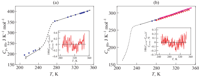
Electron microscopy and calorimetry of proteins in supercooled
Por um escritor misterioso
Last updated 16 julho 2024
Polymers, Free Full-Text

Anchored clathrate waters on the ice-nucleating sites of PbINP. A

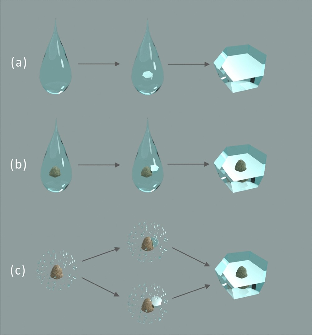
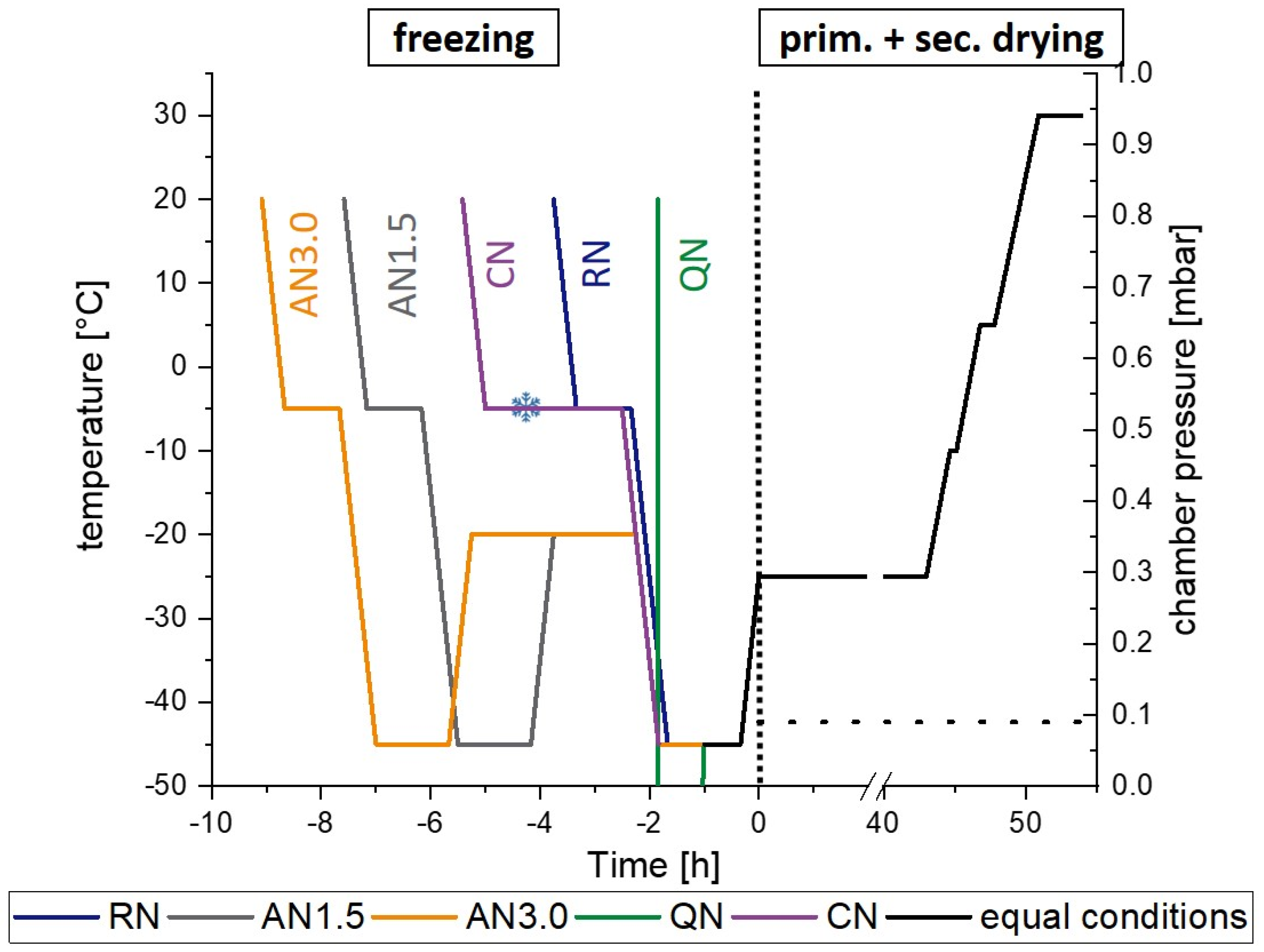
Cascade Freezing of Supercooled Water Droplet Collectives

Confined Water as Model of Supercooled Water
Polymers, Free Full-Text

Electron microscopy and calorimetry of proteins in supercooled
Electron microscopy and calorimetry of proteins in supercooled

DSC heating scans of DPPC/POPG and DPPC/PA multilamellar vesicles

a) Left: p-T diagram showing the extent of supersaturation
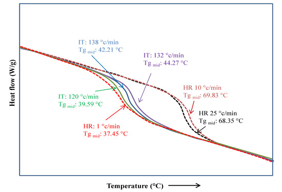
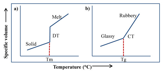
Fast Scanning Calorimetry of Organic Materials from Low Molecular

PDF) Electron microscopy and calorimetry of proteins in
Pharmaceutics, Free Full-Text
Phase change and crystallization behavior of water in biological

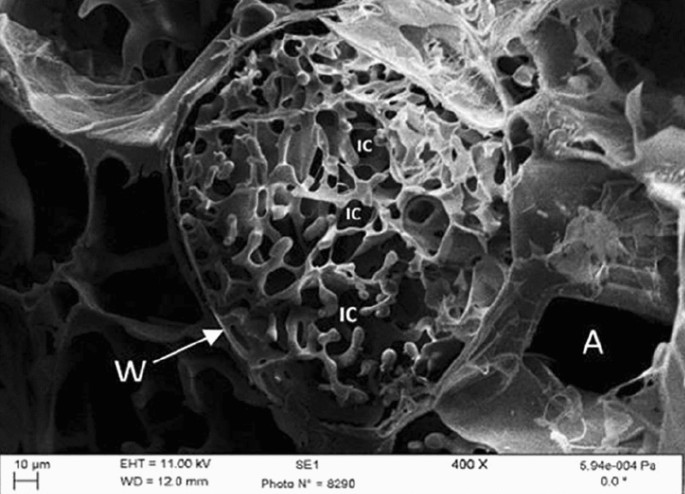
Dynamical in-situ observation of the lyophilization and vacuum
Recomendado para você
-
 WCA Live bot (unofficial) (@WCALive_bot) / X16 julho 2024
 -
 Plants, Free Full-Text16 julho 2024
 -
 Synthetic polyurethane nanofibrous membrane with sustained16 julho 2024
 -
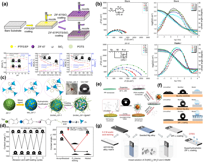 Recent progress of zeolitic imidazolate frameworks (ZIFs) in16 julho 2024
 -
Guacathon 2021 LIVE: The 21st Birthday Celebration for Joaquin16 julho 2024
-
 Space Radiobiology16 julho 2024
 -
 Methodological approach to indigenous fruit trees breeding: case16 julho 2024
 -
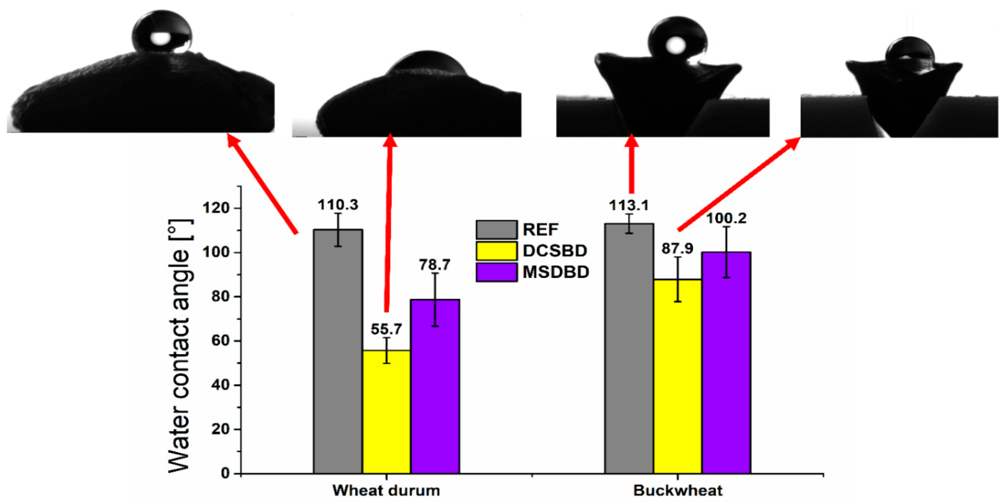
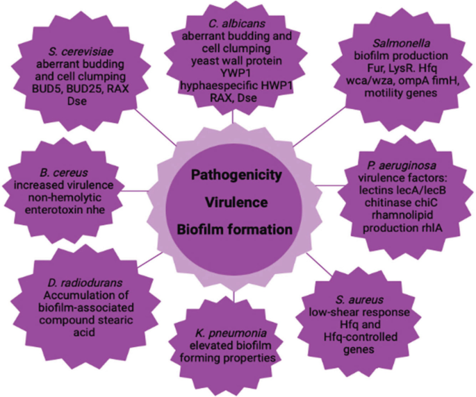
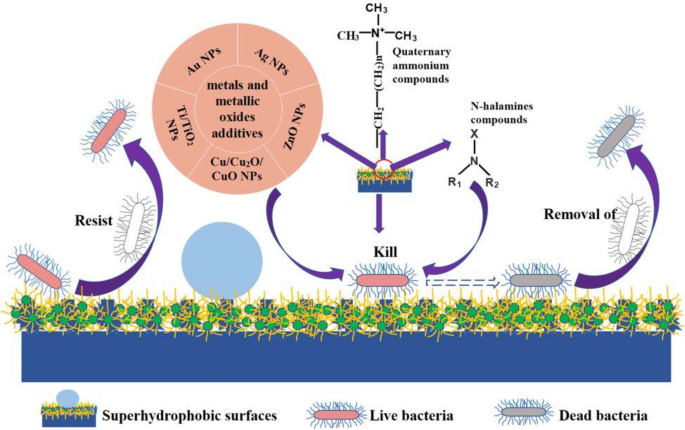 Recent Advances in Superhydrophobic and Antibacterial Cellulose16 julho 2024
 -
 Pretzel Mania 2023 World Cube Association16 julho 2024
 -
 Verified Live : BBCNEWS : October 5, 2023 3:00pm-3:31pm BST : Free16 julho 2024

você pode gostar
-
 Quest: Escape Room 2 on Steam16 julho 2024
 -
 spongebob freaking out spongebob squarepants gif16 julho 2024
 -
 LOUD apresenta novo uniforme - Game Arena16 julho 2024
 -
 B Grade ** Monitor BenQ ZOWIE 24.5 XL2566K TN FHD 360Hz DyAc+ 0.516 julho 2024
 -
 Homem morre depois de ser colhido por touro em Valência - SIC Notícias16 julho 2024
 -
 The Rock's C--, The Rock's Pancakes16 julho 2024
 -
 Minha Vez - Ton Carfi e Livinho16 julho 2024
 -
GitHub - tiptoppp/Happy-Wheels: The original Happy Wheels game in Adobe Flash16 julho 2024
-
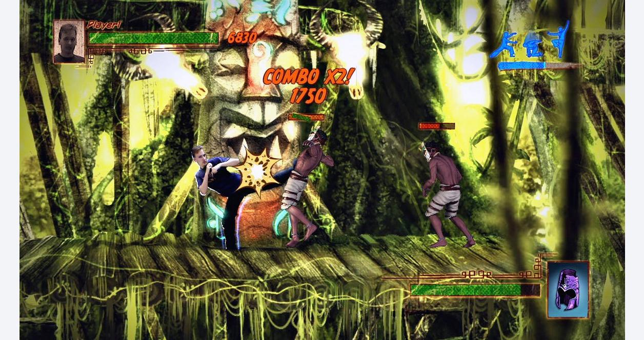
Kung Fu High Impact - Xbox 360, Xbox 36016 julho 2024
-
Twitch Lurker16 julho 2024